
Creating Effective Illustrations to Guide Students through a Dissection on the Soft- Embalmed Cadaver
ABOUT
Dissection has been viewed, both presently and historically, as an essential tool for teaching gross anatomy to health professional students, and many students in the medical and applied health fields encounter cadaveric dissection in their academic journey. Most cadavers are embalmed in a formalin-based solution, however, they do not have the same coloring, texture, flexibility, or elasticity as living tissue does, thus leaving medical and applied health students with misconceptions of the living human body. Several schools in the United States and Europe are using cadavers embalmed in a modified salt-based solution, soft-embalmed cadavers, as an addition or alternative to the formalin- embalmed cadaver.
Anatomists consider soft-embalmed cadavers to be the most accurate learning aids, more so than hard-fixed cadavers. Because soft-embalmed tissue has a unique appearance and behavior when manipulated, the illustrations in current dissection manuals insufficiently represent this tissue.
This project fulfills the visualization need of creating dissection illustrations for the soft-embalmed cadavers, increasing their accessibility in an educational environment. Moreover, this project takes advantage of the new learning opportunities that soft-embalmed cadavers can offer and aims to provide an enriched educational experience to medical and applied health students by communicating a novel approach to dissection – through a narrative of embryological development.
COMMITTEE
John Daugherty, MS
Biomedical Visualization, University of Illinois at Chicago
Christa Wellman, MFA
Dept. of Anatomy and Cell Biology, University of Illinois at Chicago
Marcelo Oliver, MFA
Body Scientific International
Noel Boaz, MD, PhD
School of Health Sciences, Emory and Henry College

Process
DISSECTION OBSERVATION AND SKETCHING
This dissection is intended for a type of cadaver that I was unfamiliar with. Thus, observation of the dissection was a critical step in producing effective visuals. I observed this dissection as it was performed by my content expert, Dr. Noel Boaz, who had been working with soft-embalmed cadavers for several years. As I was observing and partaking in this dissection, I worked to capture not only the steps of the dissection, but also the likeness of the tissue.
ESTABLISHING LEARNING GOALS AND OBJECTIVES
After carefully studying the dissection and reviewing the anatomy and embryology involved, I worked to establish the learning goals of this dissection. This allowed me to focus on a crafting a story, and target the focus of my illustrations to telling that story.
LEARNING GOAL:
Identify and understand the anatomy of the pharyngeal arch 1 derivatives on the soft-embalmed cadaver.
LEARNING OBJECTIVES:
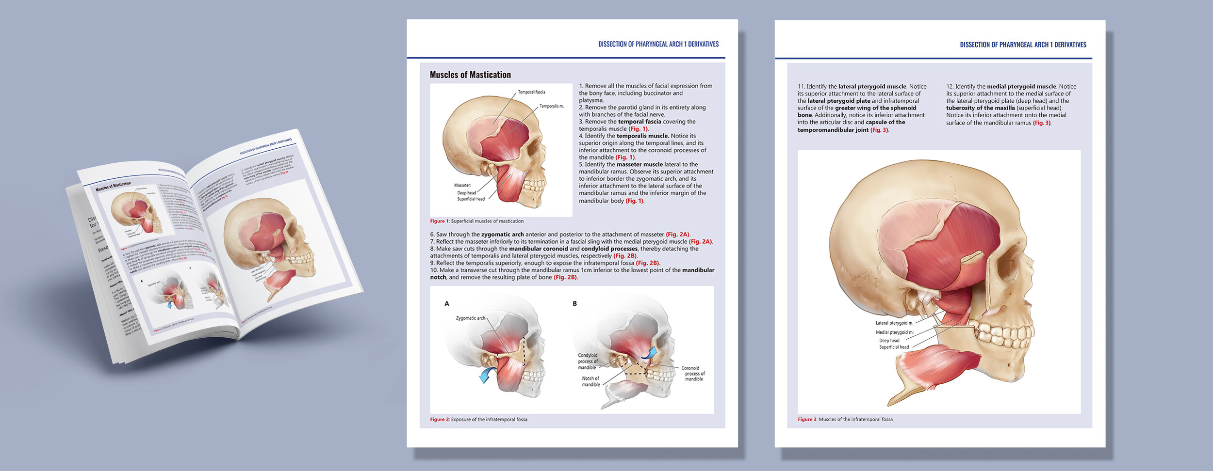
Identify the muscles of mastication, understanding their origins and insertions.
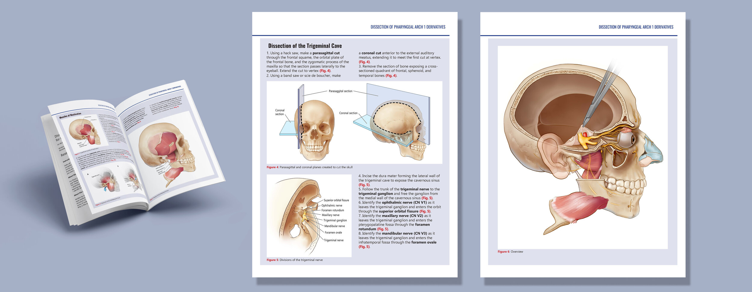
Identify the main divisions of the trigeminal nerve, understanding how they exit the skull
Identify and understand the course and branching of the ophthalmic division in the orbit.
Identify and understand the branching of the maxillary division, as well as the attachments to the pterygopalatine ganglion in the pterygopalatine fossa.
Identify and understand the course and branching of the mandibular division in the infratemporal fossa.
FROM CONCEPT TO FINAL
After defining learning goals and objectives, concept sketches were produced on paper, and agreed upon with the content expert. Then sketches were refined and rendered using Adobe Photoshop. Finally, each sketch was labeled in and designed in Adobe Illustrator. The final layout was done in Adobe InDesign.
Concept Sketch
Refined Sketch
Rendering
Design and Layout